PHILADELPHIA (June 14, 2024) — Researchers have developed a multicellular model of a pancreatic cancer tumor mass using human cells. The model uses bioprinting, which combines bioinks/biomaterials and two types of cells, to recreate the complex structure that makes pancreatic cancers among the most difficult to treat.
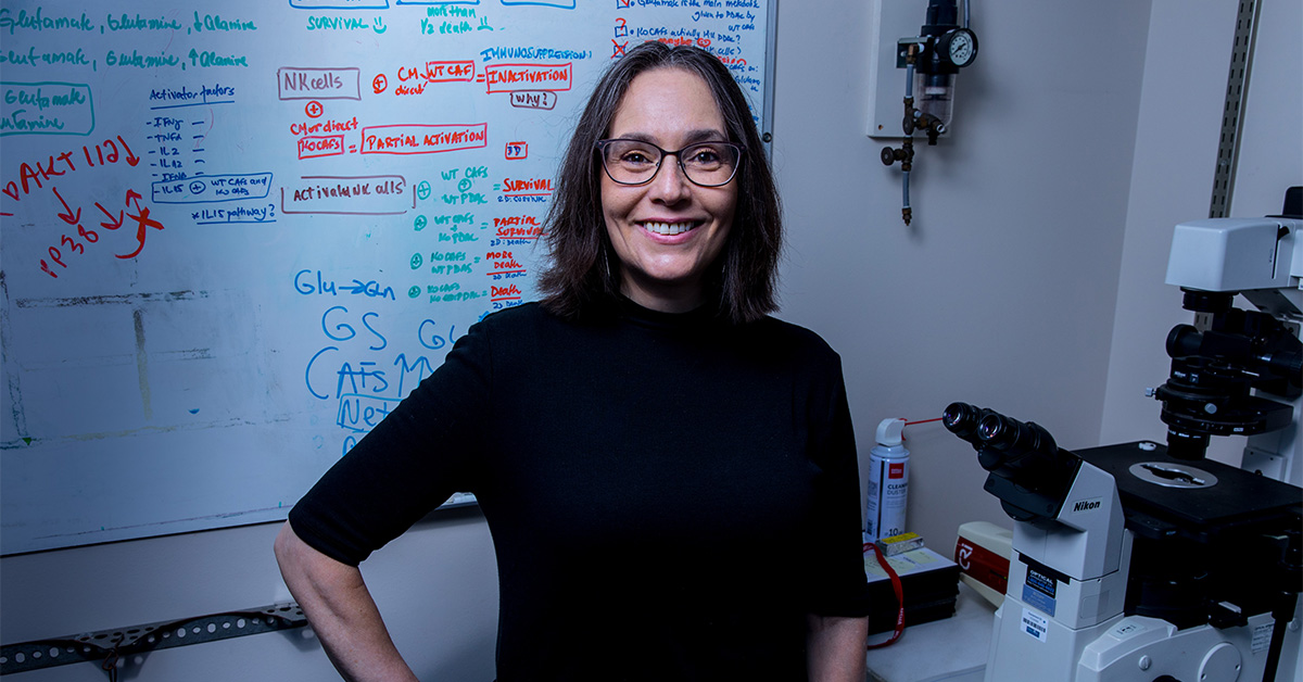
The model represents a significant advancement over previous cell models because it builds on a cellular structure that more closely mimics real human tissue, said co-corresponding author Edna Cukierman, PhD, Co-Director of the Marvin and Concetta Greenberg Pancreatic Cancer Institute and Co-Leader of the Cancer Signaling and Microenvironment Research Program at Fox Chase Cancer Center. Other Fox Chase researchers, as well as a team from Binghamton University, State University of New York, collaborated on the study.
She added that the model is a powerful tool that could be used to investigate complex cellular interactions and test novel therapies. “This will allow us to make new discoveries about how these mechanisms work with something that is in-vivo-like, three-dimensional, but also reproducible.”
Within a pancreatic cancer tumor are small “islands” of cancer cells surrounded by a large “sea” of noncancerous cells called fibroblasts. All cancers involve interactions between cancerous and noncancerous cells, but pancreatic cancer is unique because of how the fibroblasts and their self-generated extracellular matrix scaffolds far outnumber the cancer cells, ultimately overtaking the healthy pancreas with a chronically expanding scar-like tissue.
The new model discussed in the study recreates this “sea and island” structure so that researchers can study how cells interact and support each other to drive the tumor’s growth. Because the model is reproducible, it allows scientists to study what happens when they change certain conditions for the cells. In addition to using human cells, it also offers advantages over commonly used mouse studies because it allows scientists to observe cellular changes and alter selected cellular functions in real time.
Researchers have previously used organoids, artificially grown masses of cells that can mimic organ tissue, to study cancer and other diseases. However, these models rely on an externally introduced artificial scaffolding to organize cells.
For the new model, the scientists used bioink combined with human cancer-associated fibroblasts to effectively replicate the natural tumor mass scaffolding. This is an important advancement, Cukierman said, because how the cells organize and arrange themselves plays an important role in how they behave and interact.
“We felt the need to be a little more accurate in how we mimic this environment,” she said, noting that the team’s multidisciplinary background combined their expertise in bioengineering, proteomics, and the tumor microenvironment to develop a more accurate model.
The team of researchers were able to validate the model by recreating known signaling pathways between the various cells, as well as the flow of nourishment from fibroblasts to cancer cells.
“We demonstrated that it works,” Cukierman said.
She hopes to use the model to study that nourishment process further to better understand how fibroblast and their natural scaffolds functioning as a biologic unit – sea – are subverted to support cancer cells – islands – and how they could potentially be harnessed to fight it instead.
In addition to cellular experiments and drug discovery, the model could potentially be used someday to personalize cancer treatments, Cukierman noted, replicating tumor samples using a patient’s actual cells to find the therapy that is most effective.
Next, the team hopes to make the model more complex by adding more types of cells, such as endothelial and immune cells.
“This simplified model can get more complicated,” she said. “It really is just the beginning.”
The paper, “A Bioprinted Sea-and-Island Multicellular Model for Dissecting Human Pancreatic Tumor-Stroma Reciprocity and Adaptive Metabolism,” was published in Biomaterials.