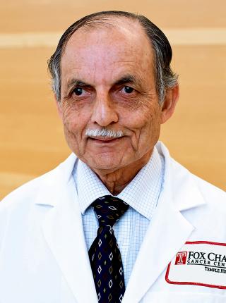
Mohan Doss, PhD, MCCPM

Mohan Doss, PhD, MCCPM

Professor

Educational Background

  • BSc, Physics, Madras University, Madras, India, 1971
  • MSc, Physics, Indian Institute of Technology, Kanpur, India, 1973
  • MS, Physics, Carnegie-Mellon University, 1975
  • PhD, Physics, Carnegie-Mellon University, 1980

Certifications

  • Member, Canadian College of Physicists in Medicine (Nuclear Medicine Physics), 1994

Honors & Awards

  • Outstanding Leadership Award in the Field of Dose Response, International Dose-Response Society, 2014
  • FCCC Ovarian SPORE Career Development Award, 2003

Research Interests

My research interest is mainly in the area of PET Imaging:

PET Imaging

Positron emission tomography (PET) with 18F-Fluorodeoxyglucose (FDG) has become an important nuclear medicine imaging technique in staging many types of cancers by providing three dimensional images of glucose metabolism. Many additional PET imaging compounds have been studied.

Research Projects

Biodistribution and Dosimetry of Novel PET Imaging Agents: When new PET imaging agents are developed to probe different biological processes, it is necessary to evaluate their safety by measuring the biodistribution of the agent and estimating the radiation dose to different organs. It is also important to know whether the normal biodistribution may obscure the anticipated biodistribution from the disease process. We have developed tools and methodology to do this evaluation using our PET/CT scanner. In a typical study, subjects are injected with the new imaging agent, and five consecutive whole body PET/CT scans are acquired over a period of about 2 hours. By examining the PET images and quantifying the uptake in the organs at the different time points, the radiation dose to the organs are estimated using the OLINDA/EXM software, and any critical organs are identified. We have studied the following F-18 labeled compounds thus far: HX-4 and VM4-037 (hypoxia imaging agents), RGD-K5 (angiogenesis marker) and W-372 (amyloid β plaque imaging agent). Studies of additional compounds are being planned.

Quantitative PET imaging of Y-90: We have investigated methods of accurately quantifying the PET images from Y-90. The PET images can be utilized for estimating the radiation dose to the tumors and organs from Y-90 microspheres which are used to treat metastatic liver cancer. This can help in optimizing the treatment. We participated in a multi-site study called QUEST, organized by one of the manufacturers of Y-90 microspheres.

Using textural analysis of PET tumor images for predicting response to treatment: In this project, we are performing textural analysis of PET images to determine some indices of heterogeneity of the activity distribution in the tumors, in order to establish a correlation between the heterogeneity indices and the subsequent response to treatment. Identifying patients with low likelihood of tumor response can help in modifying the treatments in order to achieve better outcomes.

Pre-clinical PET imaging of novel targeted drugs: We have performed pre-clinical PET imaging studies with several new targeted radiolabeled imaging agents to verify their tumor targeting.

Pre-Clinical Cerenkov imaging of novel radiotracers: In this project, we investigated the utility of Cerenkov imaging of distribution of antibody-based radiotracers in mice. This can be a very cost-effective way of determining tumor-targeting of such radiotracers.

Selected Publications

Quantitation of small-animal (124)I activity distributions using a clinical PET/CT scanner. González Trotter DE, Manjeshwar RM, Doss M, Shaller C, Robinson MK, Tandon R, Adams GP, Adler LP. J Nucl Med. 2004 Jul;45(7):1237-44. https://pubmed.ncbi.nlm.nih.gov/15235072/

Biodistribution and radiation dosimetry of the hypoxia marker 18F-HX4 in monkeys and humans determined by using whole-body PET/CT, Doss M, Zhang JJ, Belanger MJ, Stubbs JB, Hostetler ED, Alpaugh K, Kolb HC, Yu JQ. Nucl Med Comm., 31, 1016-1024 (2010). https://pubmed.ncbi.nlm.nih.gov/20948452/

Biodistribution and Radiation Dosimetry of the Integrin Marker 18F-RGD-K5 Determined from Whole-Body PET/CT in Monkeys and Humans. Doss M, Kolb HC, Zhang JJ, Bélanger MJ, Stubbs JB, Stabin MG, Hostetler ED, Alpaugh RK, von Mehren M, Walsh JC, Haka M, Mocharla VP, Yu JQ. J Nucl Med. 53, 787-795 (2012). https://pubmed.ncbi.nlm.nih.gov/22499613/

Biodistribution and radiation dosimetry of 18F-CP-18, a potential apoptosis imaging agent, as determined from PET/CT scans in healthy volunteers. Doss M, Kolb HC, Walsh JC, Mocharla V, Fan H, Chaudhary A, Zhu Z, Alpaugh RK, Lango MN, Yu JQ. J Nucl Med. 2013 Dec;54:2087-92. https://pubmed.ncbi.nlm.nih.gov/24136934/

3D inpatient dose reconstruction from the PET-CT imaging of 90Y microspheres for metastatic cancer to the liver: Feasibility study, E. Fourkal, I. Veltchev, M. Lin, S. Koren, J. Meyer, M. Doss, J. Q. Yu, Medical Physics, 40, 081702 (2013); http://dx.doi.org/10.1118/1.4810939

Biodistribution and Radiation Dosimetry of the Carbonic Anhydrase IX Imaging Agent [18F]VM4-037 determined from PET/CT Scans in Healthy Volunteers, Mohan Doss, Hartmuth C. Kolb, Joseph C. Walsh, Vani P. Mocharla, Zhihong Zhu, Michael Haka, R. Katherine Alpaugh, David Y.T. Chen, Jian Q. Yu, Molecular Imaging and Biology, 16, 739:746, 2014. http://rd.springer.com/article/10.1007%2Fs11307-014-0730-7

90Y-PET imaging: Exploring limitations and accuracy under conditions of low counts and high random fraction, Thomas Carlier, Kathy P. Willowson, Eugene Fourkal, Dale L. Bailey, Mohan Doss and Maurizio Conti, Med. Phys. 42, 4295 (2015). http://scitation.aip.org/content/aapm/journal/medphys/42/7/10.1118/1.4922685

A multicentre comparison of quantitative 90Y PET/CT for dosimetric purposes after radioembolization with resin microspheres: The QUEST Phantom Study. Willowson KP, Tapner M, The QIT, Bailey DL, Willowson KP, Tapner MJ, Ahmadzadehfar H, Amthauer H, Großer OS, Arbizu J, Martí-Climent JM, Rodriguez-Fraile M, Attarwala AA, Glatting G, Molina-Duran F, Bagni O, Filippi L, Benard F, Celler A, Bonutti F, Botta F, Ferrari M, Boucek JA, Francis R, Bourgeois AC, Bradley YC, Pasciak AS, Buchholz HG, Miederer M, Büsing KA, Schönberg SO, Carlier T, Eugene T, Cervo M, Moore SC, Civollani S, Pettinato C, Conti M, Craig AJ, Flux GD, Cremonesi M, D’Andrea M, Iaccarino G, Strigari L, D’Arienzo M, D’Asseler Y, Lambert B, Di Martino F, Doss M, Duan H, Hoffmann M, Flamen P, Vanderlinden B, Fletcher AM, Fourkal E, Freeman LM, Geatti O, Goedicke A, Habito CMR, Ouyang J, Hallam A, Morgan DG, Heard S, Leek F, Holm S, Mortensen J, de Nijs R, Hooker CA, Jeans SP, Julyan PJ, Kabasakal L, Tanyildizi H, Tanyildizi H, Kappadath SC, Siman W, Lassmann M, Schlögl S, Law MW, Lee VH, Ng SC, Lhommel R, Lodge MA, Luster M, McGowan DR, McLamb B, Kaiser HJ, Mottaghy FM, Mulder RU, Judy P, Stone JR, Lopez A, Munk OL, Staanum PF, Muzaffar R, Osman MM, Nijran KS, Towey DJ, O’Keefe GJ, Pooley RA, McKinney JM, Rausch IF, Reindl M, Sheikh A, Song N, Srinivas SM, Weir G, Yu N, Bailey DL. European Journal of Nuclear Medicine and Molecular Imaging. 2015;42(8):1202-22. http://www.ncbi.nlm.nih.gov/pubmed/25967868

Cerenkov Luminescence Imaging as a Modality to Evaluate Antibody-based Positron Emission Tomography Radiotracers. D'Souza JW, Hensley H, Doss M, Beigarten C, Torgov M, Olafsen T, Yu J, Robinson M., J Nucl Med. 2017 Jan;58(1):175-180. http://www.ncbi.nlm.nih.gov/pubmed/27539844

COVID-19 Vaccine-Related Local FDG Uptake. Doss M, Nakhoda SK, Li Y, Yu JQ. Clin Nucl Med. 2021 May 1;46(5):439-441. https://pubmed.ncbi.nlm.nih.gov/33661194/

Additional Publications

The following ratings and reviews are based on verified feedback collected from independently administered patient experience surveys. The ratings and comments submitted by patients reflect their own views and opinions. Patient identities are withheld to ensure confidentiality and privacy. Learn more about our Patient Experience Ratings.

Ratings Breakdown

Loading ...
Patient comments

Loading ...