Heinrich Roder, PhD

Heinrich Roder, PhD

This Fox Chase professor participates in the Undergraduate Summer Research Fellowship
Learn more about Research Volunteering.
Professor

Professor, Department of Biochemistry, Temple University

Adjunct Professor, Department of Biochemistry and Biophysics, University of Pennsylvania

  • Link to /sites/default/files/styles/slider_desktop/public/images/slide/6large_0.gif?itok=Br0gn6fA

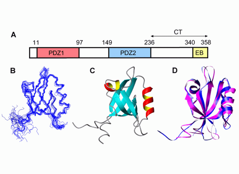
    Domain structure of NHERF1 and 3D structure of PDZ domains

  • Link to /sites/default/files/styles/slider_desktop/public/images/slide/7L_3.jpg?itok=TyRZJYDA

    Figure 7

  • Link to /sites/default/files/styles/slider_desktop/public/images/slide/8large_0.gif?itok=7JT953Zd

    Rapid formation of an intermediate during folding of the human prion protein

  • Link to /sites/default/files/styles/slider_desktop/public/images/slide/9large_1.gif?itok=e7hfLkVT

    Conformational changes associated with zymogen activation of factor XI

  • Link to /sites/default/files/styles/slider_desktop/public/images/slide/10large_0.gif?itok=V3AWDJ_K

    10 / 10Dimeric A4 dimerization domain of factor FXI

  • Link to /sites/default/files/styles/slider_desktop/public/images/slide/1large_2.gif?itok=rsukhLk3

    Structural changes in cytochrome c upon CO binding to the heme

  • Link to /sites/default/files/styles/slider_desktop/public/images/slide/2large_1.gif?itok=VCr852O8

    Kinetics of loop formation in unfolded cytochrome c

  • Link to /sites/default/files/styles/slider_desktop/public/images/slide/3large_1.gif?itok=7oVt20Ue

    Residues in staphylococcal nuclease targeted for Trp substitution

  • Link to /sites/default/files/styles/slider_desktop/public/images/slide/4large_1.gif?itok=oAdPa7n2

    Capillary mixer for ultrafast quenched-flow H/D exchange experiments

  • Link to /sites/default/files/styles/slider_desktop/public/images/slide/5large_1.gif?itok=FG2z_GX2

    Unfolding of WT apomyoglobin (left) and L115A mutant (right) at pH 4.0


    Educational Background

    • PhD, Biophysics, Swiss Federal Institute of Technology (ETH), Zürich, Switzerland, 1981

    Memberships

    • Biophysical Society
    • American Association for the Advancement of Science
    • Protein Folding Consortium
    • Protein Society
    • American Chemical Society

    Honors & Awards

    • NIH Study Section, Enabling Bioanalytical and Imaging Technology, 2018
    • NSF Panel, Mechanistic Molecular Biophysics II, 2016
    • Site Visit, NIH Bioengineering Science and Technology IRG, 2015
    • NSF Chemistry of Life Processes Panel, 2014
    • Keynote Speaker, NJACS, Princeton NJ, 2014
    • External Review Committee, Department of Biochemistry, University of Zurich, Switzerland, 2013
    • NIH NHLBI Board of Counselors, 2008
    • NIH Molecular Structure & Function B Study Section, 2008
    • Speaker and Session Chair, ACS National Meeting, Philadelphia, 2008
    • Speaker and Session Chair, Symposium on Protein Folding Dynamics, American Chemical Society National Meeting, Philadelphia, 2008
    • Editorial Board Member, Protein Engineering, Design & Selection (2004-present), Experimental Biology and Medicine (2006-present), Biophysical Journal (2007-2012)
    • Speaker and Organizer, Plenary Closing Session, 17th Symposium of the Protein Society, Boston, 2003
    • Kresge Challenge Grant and Endowment Award, 1995
    • MA, Honoris causa, University of Pennsylvania, PA, 1990

    People

    Additional Staff

    Former Staff

    Hong Cheng, PhD
    Staff Scientist

    Ruzaliya Fazlieva, MS
    Scientific Technician II

    Ming Xu, Ph.D.
    Postdoctoral Associate

    Research Interests

    Protein folding, structure and function

    • Mechanisms of protein folding, focusing on fast kinetics and early structural events
    • Protein structure, dynamics and ligand interactions, focusing on signaling adapters
    • Functional roles of intrinsically disordered protein regions
    • Methods used include NMR spectroscopy, H/D exchange, fluorescence and rapid mixing

    Lab Overview

    Our research is aimed at understanding fundamental questions in protein science, including mechanisms of protein folding, protein structure-function relationships, and the impact of protein dynamics and intrinsic disorder on protein function. My group approaches these problems using various biophysical tools, including NMR, fluorescence and other optical techniques, rapid kinetics and protein engineering.

    Lab Description

    A central theme of our research concerns the early stages of protein folding, which are critical for understanding how the native structure of a protein, and ultimately its function, are encoded in the amino acid sequence. The insight gained not only provides a basis for protein structure prediction and de novo design, but also contributes to our mechanistic understanding and treatment of a wide range of diseases that involve aggregation of denatured or misfolded proteins. The stability and folding dynamics of proteins also have major implications with respect to understanding of the physiological consequences of mutations, in vivo folding and other cellular processes, such as trafficking and degradation. We study the dynamics of protein folding on a microsecond time scale by coupling advanced mixing techniques with various detection methods, including fluorescence and H/D exchange labeling experiments with NMR detection. In combination with protein engineering, these approaches have provided detailed insight into the folding mechanisms of a diverse set of proteins.

    A recent effort has been to elucidate the mechanism of coupled protein folding/ligand binding reactions. We explored the ligand-induced folding reaction of staphylococcal nuclease (SNase) under conditions (2.9 M Urea) where the free protein is unfolded, but the complex with a nucleotide analog (prAp) is folded, using optically monitored kinetic measurements, NMR and molecular dynamics simulations. Theoretical modeling of the kinetic data using statistical mechanics showed that the conformational changes precede binding at low ligand concentrations (“conformational selection”), whereas the second-order binding step preceded formation of the native structure at high ligand concentrations (“induced fit”). Real-time 1D and 2D NMR measurements confirmed that an encounter complex of prAp with a partially structured, but dynamic, form of SNase accumulates transiently at high ligand concentrations. The results shed new light on the structural/energetic basis of coupled folding/binding reactions, which are highly relevant not only for understanding molecular recognition in cases where at least one of the binding partners is intrinsically disordered.

    Our group also investigates the structure, dynamics and molecular interactions of various proteins of biomedical interest in solution by using NMR spectroscopy and other biophysical methods. An ongoing project is aimed at understanding the structural and dynamic properties of Na+/H+ exchanger regulatory factor (NHERF), a signaling protein comprising two PDZ domains and a C-terminal ezrin binding motif, as well as two long intrinsically disordered regions. Detailed NMR and thermodynamic studies have shown that the activity of NHERF as a signaling adaptor at the membrane-cytoskeleton interface is regulated by a complex equilibrium between various open and closed conformations. By combining our biophysical approaches with cellular imaging and flow cytometry, we have gained detailed insight into the mechanism by which NHERF regulates the signaling activity and trafficking of the epidermal growth factor receptor (EGFR).

    More broadly, we are interested in understanding how interactions between multivalent binding partners, such as NHERF or the SH2-domain containing phosphatase Shp2, and intrinsically disordered cytosolic regions of membrane receptors, such as EGFR or the killer cell inhibitory receptor KIR3DL, can mediate receptor clustering and phase separation on the plasma membrane. To resolve and assign the crowded NMR spectra of these intrinsically disordered protein regions (IDRs) we make extensive use of 13C-detected NMR techniques, including a novel technique for mapping recognition sites for globular binding partners on IDRs. By applying these approaches to the cytosolic tail of KIR3DL1, we gained detailed insight into the secondary structure propensities of this 84-residue IDR, membrane-interacting regions, and binding sites for a key interaction partner, the phosphatase Shp2, which recognizes a pair of phospho-tyrosine sequence motifs via a pair of SH2 domains. These findings provide a structural basis for understanding how interactions between two bivalent binding partners regulate receptor signaling.

    Misc

     
    Former Staff

    Ming Xu

    Software

    Sphere: A server program for hydrogen exchange rate estimation

    Selected Publications

    Mizukami T., Bedford J.T., Liao S., Greene L.H., Roder H., Effects of ionic strength on the folding and stability of samp1, a ubiquitin-like halophilic protein. Biophys J. 121(4): 552-564, 2022. PMC8874027. https://www.ncbi.nlm.nih.gov/pubmed/35063455.

    Mizukami, T.; Furuzawa, S.; Itoh, S. G.; Segawa, S.; Ikura, T.; Ihara, K.; Okumura, H.; Roder, H.; Maki, K., Energetics and Kinetics of Substrate Analog-Coupled Staphylococcal Nuclease Folding Revealed by a Statistical Mechanical Approach. Proc. Natl. Acad. Sci. U. S. A. 2020; 117: 19953-19962. PMC7443883

    Iwafuchi, M.; Cuesta, I.; Donahue, G.; Takenaka, N.; Osipovich, A. B.; Magnuson, M. A.; Roder, H.; Seeholzer, S. H.; Santisteban, P.; Zaret, K. S., Gene Network Transitions in Embryos Depend Upon Interactions between a Pioneer Transcription Factor and Core Histones. Nat Genet 2020; 52: 418-427.

    Cheng, H.; Schwell, V.; Curtis, B. R.; Fazlieva, R.; Roder, H.; Campbell, K. S., Conformational Changes in the Cytoplasmic Region of Kir3dl1 Upon Interaction with Shp-2. Structure 2019; 27: 639-650 e2. PMC6447435

    Bai, N.; Roder, H.; Dickson, A.; Karanicolas, J., Isothermal Analysis of Thermofluor Data Can Readily Provide Quantitative Binding Affinities. Sci Rep 2019; 9: 2650. PMC6389909

    Cheng, H.; Roder, H. Carbon-Detected NMR for Mapping Binding Sites in Intrinsically Disordered Regions of a Protein. US Patent No. 10,436,795 B2, 10/8/2019, 2019.

    Mizukami, T.; Xu, M.; Fazlieva, R.; Bychkova, V. E.; Roder, H., Complex Folding Landscape of Apomyoglobin at Acidic Ph Revealed by Ultrafast Kinetic Analysis of Core Mutants. J Phys Chem B 2018; 122: 11228-11239. PMC6395567

    Alves, C.; Cheng, H.; Tavanez, J. P.; Casaca, A.; Gudima, S.; Roder, H.; Cunha, C., Structural and Nucleic Acid Binding Properties of Hepatitis Delta Virus Small Antigen. World J Virol 2017; 6: 26-35. PMC5437381

    Rawat, S. J.; Araiza-Olivera, D.; Arias-Romero, L. E.; Villamar-Cruz, O.; Prudnikova, T. Y.; Roder, H.; Chernoff, J., H-Ras Inhibits the Hippo Pathway by Promoting Mst1/Mst2 Heterodimerization. Curr Biol 2016; 26: 1556-63. PMC4915977

    Wu, Y.; Span, L. M.; Nygren, P.; Zhu, H.; Moore, D. T.; Cheng, H.; Roder, H.; DeGrado, W. F.; Bennett, J. S., The Tyrosine Kinase C-Src Specifically Binds to the Active Integrin Alphaiibbeta3 to Initiate Outside-in Signaling in Platelets. J. Biol. Chem. 2015; 290: 15825-34. 4505490

    Lehmann, A.; Wixted, J. H.; Shapovalov, M. V.; Roder, H.; Dunbrack, R. L., Jr.; Robinson, M. K., Stability Engineering of Anti-Egfr Scfv Antibodies by Rational Design of a Lambda-to-Kappa Swap of the Vl Framework Using a Structure-Guided Approach. mAbs 2015; 7: 1058-71. PMC4966335... Expand

    Additional Publications

    The following ratings and reviews are based on verified feedback collected from independently administered patient experience surveys. The ratings and comments submitted by patients reflect their own views and opinions. Patient identities are withheld to ensure confidentiality and privacy. Learn more about our Patient Experience Ratings.

    Ratings Breakdown

    Loading ...

    Patient comments

    Loading ...

    This Fox Chase professor participates in the Undergraduate Summer Research Fellowship
    Learn more about Research Volunteering.