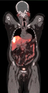
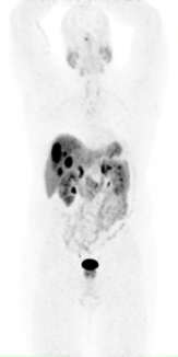
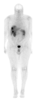
Neuroendocrine tumors are often found accidentally before they cause any symptoms, often when a patient is being evaluated for another condition, such as iron deficiency anemia.
Conventional imaging techniques such as CT, FDG PET, ultrasound, or MRI are often not able to detect NETs early because of the small size of the tumor, low metabolic rate and irregular location in the body.
But, Fox Chase Cancer Center was the first hospital in the greater Philadelphia and tri-state region to introduce a new FDA-approved imaging method called Gallium68 DOTATATE PET/CT that allows for much greater precision than ever before with a much higher rate of detection.
Gallium68
The Gallium68 PET scan is PET and CT used simultaneously, with PET capturing images of organ and tissue function and CT capturing body structure. The integrated imaging machines use a “tracer” (Ga68 DOTATATE) injected into the patient to highlight areas of the body where cancer is present. Hundreds of images are taken per session in three dimensions, which stack up to form a patient’s whole body display. This allows the radiologists and physicians at Fox Chase to examine and evaluate a patient one portion at a time in great detail.
In addition to providing excellent image quality, the Gallium68 PET scan takes much less time than routine nuclear medicine imaging (such as the OctreoScan). Instead of a series of scans over three days, the Gallium68 PET scan takes about three hours, and patients are exposed to much less radiation compared to older methods.
Along with being used for diagnosis, the scan is also useful for follow up on treatment response. The Ga68 PET scan is also used to select patients for the recently FDA-approved peptide receptor radiopharmaceutical therapy (PRRT).


